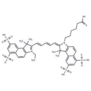
| Name | Cy5.5 |
| Description | Cy5.5 is a near-infrared fluorescent dye for labeling biomolecules such as peptides, proteins and oligonucleotides (Ex=673 nm, Em=707 nm)[2]. |
| In vitro | I. Preparation of stock solution steps
1. Protein preparation
1) To achieve the best labeling effect, the protein (antibody) concentration needs to be adjusted to 2 mg/mL.
2) Ensure that the pH of the protein solution is within the range of 8.5±0.5. If the pH value is lower than 8.0, 1 M sodium bicarbonate can be used to adjust.
3) When the protein concentration is lower than 2 mg/mL, the labeling efficiency will be significantly reduced. To optimize the labeling effect, it is recommended that the protein concentration be maintained between 2-10 mg/mL.
4) The protein needs to be dissolved in a buffer that does not contain primary amines (such as Tris or glycine) and ammonium ions, otherwise it will interfere with the labeling reaction.
2. Dye preparation
Use anhydrous DMSO to prepare a 10 mM stock solution of Cy5.5 dye. Mix well with a glass rod or vortex.
Note: It is recommended that the Cy5.5 dye stock solution be stored at -20 ℃ or -80 ℃ in the dark after aliquoting. Before subsequent labeling experiments, 500 μg/mL Activate with condensation solution (EDC hydrochloride T19947).
3. Calculation of dye working solution dosage
The amount of Cy5.5 dye used depends on the amount of labeled protein. The optimal molar ratio of Cy5.5 dye to protein is about 10.
Example: 500 μL, 2 mg/mL IgG (MW = 150,000) needs to be labeled. Assuming that 100 μL DMSO is used to dissolve 1 mg of CY dye, the calculation is as follows:
1) IgG (mmol) = IgG (mg/mL) ×IgG (mL) / IgG (MW) =2 mg/mL×0.5 mL / 150000 mg/mmol= 6.7×10^6 mmol
2) Cy5.5 (mmol) = IgG (mmol) ×10 =6.7×10^6 mmol×10 = 6.7×10^5 mmol
3) Cy5.5 (μL) = Cy5.5 (mmol)×Cy5.5 (MW)/mg/Cy5.5 (μL) = 6.7×10^5 mmol× 917.06 g/mol / 0.01 mg/μL
II. Labeling reaction
1) Take the calculated amount of freshly prepared 10 mM Cy5.5 dye master solution (about 10 μL) and add 50 μL of 500 μg/mL condensation solution for activation. Slowly add this mixture to 0.5 mL of protein sample solution, gently mix and briefly centrifuge to allow the sample to sink to the bottom of the reaction tube. Avoid violent shaking to prevent protein denaturation or inactivation.
2) Place the reaction tube in a light-proof environment and gently shake and incubate at room temperature for 60 minutes. Every 10-15 minutes, gently flip the reaction tube several times to ensure adequate mixing and improve labeling efficiency.
The above information is based on published literature. Experimental procedures should be appropriately modified to meet specific research demands. |
| In vivo | METHODS: Cy5.5-FFRck-fVIIa and Cy5.5 dye alone were injected intravenously (i.v.) into right flank athymic nude mice (nu/nu) bearing TF-expressing ASPC-1 and TF-non-expressing MiaPaCa pancreatic tumor xenografts and tested for duration and stability of binding to enable follow-up of treatment response.
RESULTS Specific localization of Cy5.5-FFRck-fVIIa to TF in VECs of pancreatic tumor xenografts was observed from day 1 to day 26 in both ASPC-1 and MiaPaCa tumors, whereas no specific localization was observed with unconjugated Cy5.5 alone.[1] |
| Storage | Powder: -20°C for 3 years
Shipping with blue ice/Shipping at ambient temperature. |
| Solubility Information | H2O : 6.7 mg/mL (7.31 mM), The compound is unstable in solution. Please use soon.
DMSO : 85 mg/mL (92.69 mM), The compound is unstable in solution. Please use soon.
10% DMSO+40% PEG300+5% Tween-80+45% Saline : 5 mg/mL (5.45 mM), Sonication is recommended.
|
| Keywords | Inhibitor | inhibit | Cy-5.5 | Cy5.5 | 210892-23-2 |

 United States